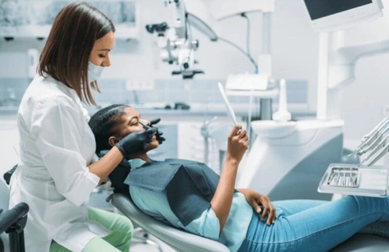

Mohs surgery is a specialized surgical technique often recommended for treating certain types of skin cancer. The procedure has become a well-regarded method for removing cancerous tissue while preserving as much healthy skin as possible. A unique aspect of Mohs surgery is the role pathology plays throughout the process. While patients may hear terms like “margins” and “microscopic examination” during discussions with Mohs surgeons, understanding how pathology intersects with this surgical procedure can provide a clearer picture of what to expect.
What Mohs Surgery Is?
Mohs surgery is a precise, layer-by-layer approach to excising cancerous tissue. It is often recommended for skin cancers that appear in cosmetically or functionally sensitive areas, such as the face. The procedure is conducted in stages and is usually performed under local anesthesia.
Mohs surgeons focus on conserving as much non-cancerous tissue as possible. Each stage involves removing a thin layer of skin, which is then examined under a microscope for the presence of cancer cells. If cancer is found at the edges of the examined tissue, additional layers are removed and examined until no remaining cancer is detected. This highly meticulous process enables the surgeon to map out the extent of the cancer and achieve a high level of accuracy in its removal.
Pathology’s Role in Mohs Surgery
Pathology is a core component of Mohs surgery, seamlessly integrated into the procedure to guide the accurate removal of cancerous tissue. During the surgery, the surgeon also acts as a pathologist. They examine the excised layers of skin to determine whether cancer cells are present during the surgery procedure.
Microscopic Examination of Skin Samples
Each sample removed during Mohs surgery is carefully processed and analyzed. After a layer of skin is excised, it is immediately frozen, thinly sliced, and placed onto microscope slides for examination. The surgeon uses a microscope to check the sample to assess whether any cancerous cells remain. If cancer cells are detected, the surgeon has a map indicating the exact location of the remaining cancer. This means that in subsequent stages, only the areas harboring cancer are removed, leaving healthy tissue untouched.
Accuracy in Mapping Tumor Margins
Using both surgical precision and pathological expertise, the surgeon creates a detailed map of the tumor and surrounding tissue throughout the procedure. This mapping process is a distinctive feature of Mohs surgery. The pathology results are used to guide the next steps, enabling the surgeon to target cancerous areas with exceptional accuracy. This combination of surgical and pathological skills makes sure that all cancer cells are removed, significantly lowering the likelihood of recurrence. It also minimizes the removal of healthy tissue, preserving the function and appearance of the affected area.
Also Read: Ways to Prepare for Mohs Surgery
Seeking Mohs Surgeons
For patients diagnosed with skin cancer, looking at treatment options can feel like an overwhelming process. Mohs surgery often stands out as a preferred approach due to its precision and unique integration of pathology. When selecting a Mohs surgeon, it may be helpful to seek professionals with specialized training in both dermatology and Mohs procedures. Those seeking further clarity about the procedure are encouraged to discuss it with a qualified surgeon to better understand the process and whether it is well-suited to their specific needs.